Heart Disease – Coronary Disease
Mitral Valve Surgery
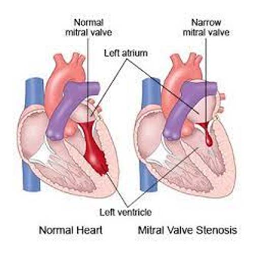
Blood is difficult to pass from the left atrium to the left ventricle due to narrowing of the mitral valve. The amount of blood that accumulates in the left atrium can causes it to swell up. These causes irregular heartbeats (palpitations) – Atrial fibrillation. The blood further backlogs into the lungs where it can cause raised blood pressure in the lungs (pulmonary hypertension), water accumulation and breathlessness.
Mitral stenosis is most often caused by Rheumatic Fever which may have been caught and recovered from many years beforehand. Rheumatic Fever is an inflammatory disease that occurs in a small number of patients after throat infections caused by a bacteria called streptococcus pyogenes. Now that understanding of and treatments for this are better understood, it is rare to develop Rheumatic Fever in the Singapore.
Patient have the following symptoms
Further management
Mild disease can be treated symptomatically with water tablets (diuretics). Moderate to severe disease requires mechanical correction of the valve obstruction by valvotomy, valve repair, or valve replacement.
Irregular heart beats (atrial fibrillation) causes clot formation in the heart which can lead to stroke or clots in the blood including the legs. Anticoagulation (strong blood thinners) are required to manage this as a preventive measure.
Mitral Valve Anatomy
The heart is divided into two sides; right and left.
How the heart works?
Your heart is roughly the size of a fist and sits in the middle of your chest, slightly to the left.
The Heart
It is the best pump in the world which powers your body. It supplies blood carrying oxygen and nutrients to every vital organ in your body. The heart does this all day and all night, all the time. It is located in your chest between your lungs, more to the left, and is protected by your breast bone and ribs. The heart is around the size of your clenched fist.
The heart has two sides; left and right. All the blood returns to the right side and then it is pumped to the lungs. The oxygenated blood returns to the left side of the heart and is pumped out to the vital organs.
The heart is made up of four different zones, two on each side. One is on the top and one is on the bottom. The ones on top are the atria. The heart has a left atrium and a right atrium. The two on the bottom are called the ventricles. The heart has a left ventricle and a right ventricle. The atria and ventricles work as a team.
The left side of the heart delivers oxygenated blood to vital organs. The organs takes in the oxygen and nutrients out of the blood then sends to blood filled with waste products (carbon dioxide and others) to the right side of the heart. The right ventricle pumps the blood to the lungs for oxygenation and removal of carbon dioxide. This continues over and over again.
Blood flow is like the road traffic. It should only flow in one direction. Road traffic is managed by traffic lights and blood flow is managed by valves. Valves are like doors. Doors have a frame and some have two or three doors. Valves are similar. Valves are between atria and ventricles; tricuspid (right) and mitral (left). There are two more valves; between the right ventricle and pulmonary artery (pulmonary valve) and between the left ventricle and aorta (aortic valve).
Valves can have problems like traffic lights. They can stop working the way they should. Valves can be too tight for blood to enter (stenosis) or too lax that blood backflows (regurgitation).
In summary the heart has 4 valves:
- Aortic valve – Left – between the left ventricle and aorta
- Mitral valve – Left – between the left atrium and left ventricle
- Tricuspid valve – Right – between the right atrium and right ventricle
- Pulmonary valve – Right – between the right atrium and pulmonary (lung) artery

